
In today’s fast-paced healthcare environment, precision, speed, and collaboration are vital. That’s why IICS Technologies Limited brings you a groundbreaking health software product — Radiology Information System (RIS) integrated with Picture Archiving & Communication System (PACS) in 2D & 3D. This innovative system is transforming how healthcare professionals manage, interpret, and store diagnostic imaging, leading to better outcomes for patients and improved efficiency for healthcare providers.
What is RIS with PACS in 2D & 3D?
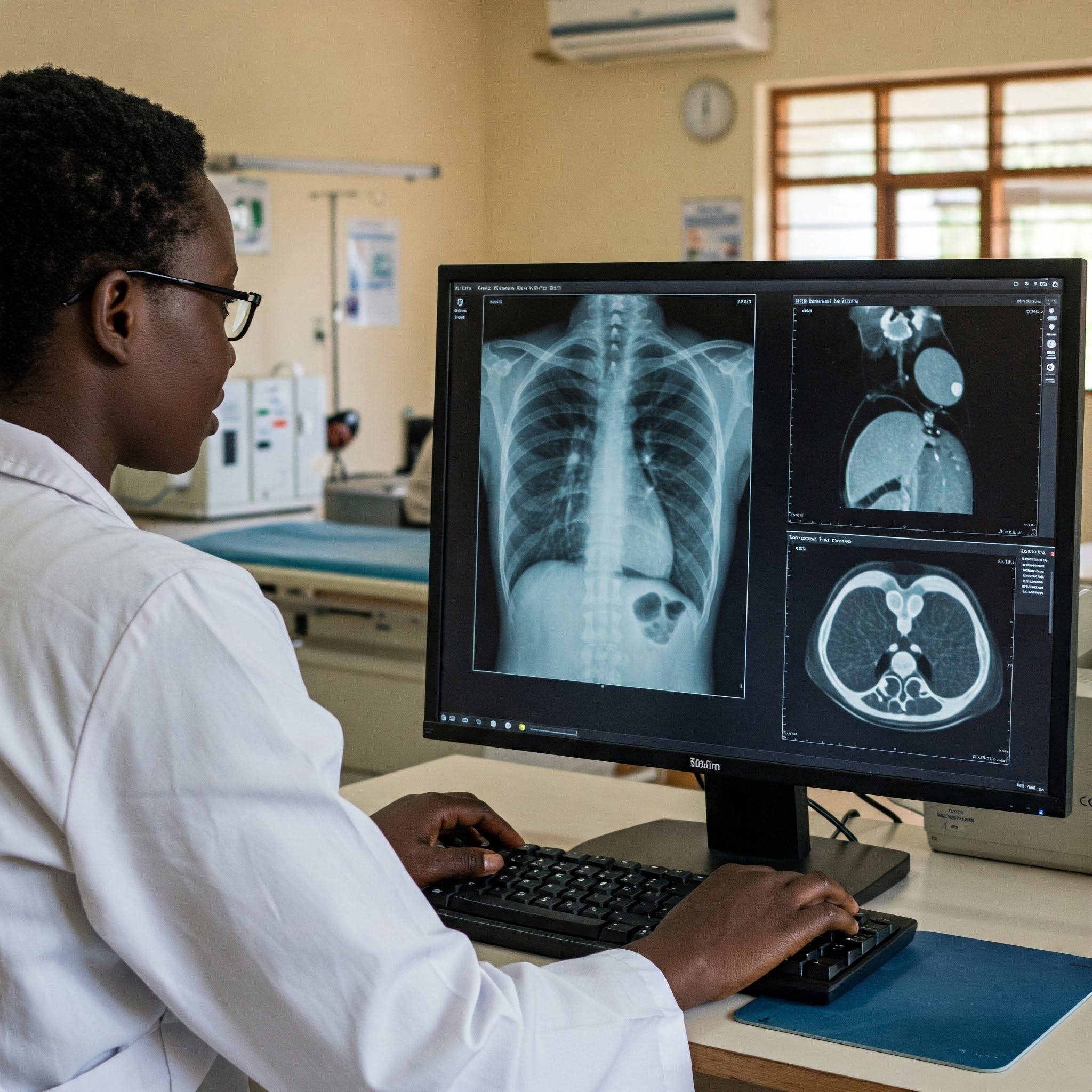
RIS with PACS is a comprehensive, end-to-end digital solution that manages radiological imaging workflows from scheduling and reporting to image archiving and distribution. The integration of 2D and 3D imaging elevates this system beyond traditional platforms, enabling sharper, more accurate diagnostics and deeper clinical insights.
-
RIS (Radiology Information System): Handles administrative and clinical tasks such as patient registration, image tracking, appointment scheduling, and results reporting.
-
PACS (Picture Archiving and Communication System): Digitally stores, retrieves, manages, and shares medical images, accessible from any authorized device or location.
-
2D & 3D Imaging: Delivers multi-dimensional views that allow radiologists and specialists to visualize internal organs, tissues, and abnormalities with exceptional clarity and depth.
Key Diagnostic Applications
RIS with PACS is used to diagnose a wide range of conditions across various medical specialties. Some of the major diseases and conditions commonly diagnosed using this system include:
1. Cancer
-
Breast Cancer: Detected using digital mammography integrated into the RIS, where 3D tomosynthesis reveals hidden tumors even in dense breast tissue.
-
Lung Cancer: Identified through CT scans with 3D reconstructions that help pinpoint nodule size, shape, and exact location in the lungs.
-
Brain Tumors: MRI images managed via PACS are layered in 3D to map tumor boundaries and assess pressure on brain structures.
2. Cardiovascular Diseases
-
Coronary Artery Disease: Diagnosed with 3D CT angiography visualized in PACS, highlighting plaque buildup in arteries with contrast imaging.
-
Congenital Heart Defects: Pediatric cardiologists use RIS to access cross-sectional 3D echo images for early diagnosis and surgical planning.
-
Aneurysms: 3D volume-rendered CT scans track blood vessel walls, allowing precise detection of aneurysm size and risk of rupture.
3. Neurological Disorders
-
Stroke: Swift diagnosis using high-resolution MRI sequences stored in PACS, showing areas of brain ischemia in 2D slices and 3D overlays.
-
Multiple Sclerosis: Tracked over time through RIS-scheduled MRI scans, identifying lesions in the brain and spinal cord using contrast enhancement.
-
Spinal Cord Injuries: RIS coordinates real-time access to 3D spinal reconstructions from CT scans, helping surgeons plan treatment interventions.
4. Orthopedic Conditions
-
Fractures and Joint Dislocations: Diagnosed using high-definition X-rays; RIS tracks patient recovery through follow-up images.
-
Arthritis and Osteoporosis: Bone density is analyzed through DEXA scans stored in PACS; 3D rendering visualizes joint erosion.
-
Post-surgical Follow-ups: RIS manages scheduled reviews with 3D orthopedic scans to monitor prosthesis placement and bone healing.
5. Pulmonary Disorders
-
Tuberculosis, COPD, Pneumonia: Detected via chest X-rays and CT scans; PACS enables comparative viewing of lung fields for disease progression.
-
COVID-19 Complications: Monitored with serial CT imaging in RIS, mapping lung inflammation in 3D to assess severity and treatment response.
-
Lung Function Analysis: PACS supports dynamic 3D airflow simulations for pulmonologists to evaluate airway obstructions or restrictions.
6. Abdominal and Pelvic Diseases
-
Liver Cirrhosis: CT scans stored and analyzed through PACS highlight liver surface irregularities and portal vein changes.
-
Kidney Stones: Identified in 2D/3D CT scans with PACS-assisted measurement tools showing size, location, and risk of obstruction.
-
Uterine Fibroids: Ultrasound images tagged and tracked in RIS offer 3D visualization of fibroid size and position, aiding gynecological decisions.
Why Choose IICS Technologies’ RIS with PACS?
-
Seamless Integration with other hospital systems like EMR/HMIS
-
Secure & Scalable Architecture that supports remote access
-
AI-ready Interface for future upgrades and predictive diagnostics
-
Customizable Workflows tailored to hospital and clinic needs
-
Cost-Efficient & Paperless solution for modern healthcare environments
Experience Precision Healthcare Today
From early detection to post-treatment evaluation, our RIS with PACS in 2D & 3D is helping hospitals, clinics, and diagnostic centers across Africa deliver world-class care. Let IICS Technologies be your partner in digital transformation.
Are You Ready to Modernize Your Radiology Department?
Contact IICS Technologies Limited today for a free demo and discover how our Radiology Information System with PACS can revolutionize diagnostics at your facility.
Contact us here to book a demo or live chat right now for more info!

